Transesophageal Echocardiography (TEE)
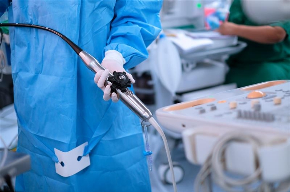
Transesophageal Echocardiography (TEE) in Patna
Advanced Heart Imaging for Accurate Cardiac Diagnosis
Transesophageal Echocardiography (TEE) is a specialized cardiac imaging test that provides detailed and high-resolution views of the heart using an ultrasound probe passed through the food pipe (esophagus). Since the esophagus lies close to the heart, TEE offers clearer images of heart chambers, valves, and major blood vessels compared to a standard echocardiogram in certain situations.
At Dr. Ashish Kumar’s cardiac care practice in Patna, TEE is performed to help diagnose complex heart conditions with greater accuracy and guide appropriate treatment decisions. The procedure supports detailed evaluation of heart structures and helps cardiologists assess conditions that may require advanced cardiac management.
What is a TEE Scan?
A Transesophageal Echocardiography (TEE) scan is a minimally invasive ultrasound-based cardiac test that captures detailed images of the heart from inside the esophagus. Unlike a regular 2D echocardiogram performed from the chest wall, TEE places the ultrasound probe closer to the heart, allowing better visualization of areas that may be difficult to assess through conventional imaging.
This advanced diagnostic technique helps evaluate heart valves, blood flow patterns, clots, infections, and structural abnormalities with improved clarity.
Why is TEE Recommended?
A cardiologist may recommend TEE when a detailed assessment of the heart is required or when standard echocardiography does not provide sufficient information.
TEE may be advised for:
- Detailed evaluation of heart valves
- Detection of blood clots inside the heart chambers
- Assessment of valve-related diseases
- Evaluation of structural abnormalities of the heart
- Diagnosis of certain congenital heart conditions
- Monitoring during specific cardiac procedures
- Assessment of heart function after certain cardiac events
How is a TEE Scan Performed?
Before the procedure, the patient’s throat is usually numbed, and medication may be provided to help with relaxation and comfort. A flexible ultrasound probe is gently guided through the mouth into the esophagus.
The probe captures detailed images of the heart, including:
- Heart chambers
- Heart valves
- Major blood vessels
- Blood flow patterns
- Internal heart structures
The procedure is generally completed within a short duration, and patients are monitored before and after the scan for safety.
Benefits of Transesophageal Echocardiography
TEE provides several advantages in advanced cardiac evaluation, including:
- High-quality images of internal heart structures
- Better visualization of heart valves and chambers
- Improved detection of small abnormalities
- Accurate assessment for treatment planning
- Useful guidance during cardiac procedures
Conditions Evaluated Through TEE
TEE can help assess various cardiovascular conditions, such as:
- Heart valve disorders
- Infective endocarditis
- Blood clots in the heart
- Atrial septal defects and other structural heart problems
- Aortic diseases
- Certain causes of stroke related to cardiac conditions
TEE Scan by Dr. Ashish Kumar in Patna
Dr. Ashish Kumar, Consultant Cardiologist at Mediversal Hospital, Patna, provides advanced cardiac evaluation with a focus on accurate diagnosis and personalized treatment planning. With expertise in non-interventional and interventional cardiology, he uses modern diagnostic approaches like TEE to understand complex heart conditions and deliver appropriate cardiac care.
If you have been advised a TEE scan or require detailed evaluation of your heart health, consult a cardiology specialist for expert guidance.